Accurate medical imaging is an important part of modern healthcare, especially when doctors need detailed information about suspicious tissues or possible cancer. Advanced imaging technologies can provide both structural and functional information about the body. PET and CT are often combined to create a comprehensive examination that can support diagnosis, staging, treatment planning, and follow-up.
Understanding PET CT Imaging
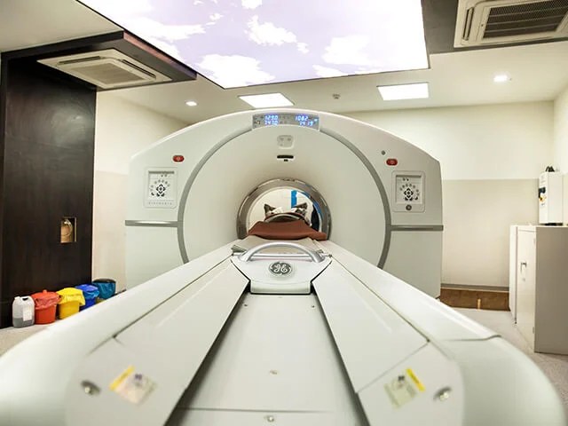
PET CT imaging combines Positron Emission Tomography (PET) and Computed Tomography (CT) in one examination. CT produces detailed anatomical pictures of organs and tissues, while PET provides information about metabolic activity within the body.
Before the scan, patients are generally given a small amount of radioactive tracer. The tracer travels through the body and may accumulate in tissues with increased metabolic activity. The PET component detects this activity, while CT helps identify the exact location and structure of the area being evaluated.
How PET CT Helps Doctors
The combination of metabolic and anatomical information can provide a more complete view than either imaging method alone in suitable clinical situations. PET can highlight areas with unusual activity, while CT shows their physical characteristics and location.
This information may help specialists evaluate suspicious lesions, assess the extent of disease, and determine whether additional investigations are necessary. Results are interpreted alongside the patient’s medical history, symptoms, laboratory findings, and other diagnostic tests.
PET CT for Cancer Diagnosis
PET CT for cancer diagnosis can be useful when doctors are evaluating a suspected or confirmed malignancy. The examination may help identify metabolically active abnormalities and provide information about whether disease appears localized or may involve multiple parts of the body.
PET CT can be used in the evaluation of selected cancers, including certain lung cancers, lymphomas, colorectal cancers, breast cancers, and head and neck cancers. Its usefulness depends on the specific cancer, clinical circumstances, and tracer being used.
Does PET CT Replace a Biopsy?
Although PET CT provides valuable imaging information, it does not necessarily replace a biopsy. In many situations, tissue examination is needed to confirm whether a suspicious lesion is cancerous and to identify its specific characteristics.
Doctors may use PET CT findings to complement biopsy results and other investigations. A complete diagnosis is based on the overall clinical picture rather than a single scan or test.
PET CT and Cancer Staging
Once cancer has been diagnosed, staging helps doctors determine how extensively the disease has developed. Knowing whether cancer is confined to the primary location or has potentially spread to lymph nodes and distant organs can influence treatment planning.
PET CT can examine several areas of the body and may identify regions showing abnormal tracer uptake. This information can help specialists evaluate the possible distribution of disease and determine whether further testing is appropriate.
Monitoring Treatment Response
PET CT may also have a role in monitoring cancer treatment. Depending on the patient’s diagnosis and treatment plan, doctors may recommend follow-up imaging after a particular period of therapy.
Comparing current images with previous scans can help specialists assess changes in metabolic activity and anatomical appearance. Changes in tracer uptake may provide useful information about treatment response, although results should always be interpreted alongside clinical findings.
Preparing for PET CT
Preparation requirements vary according to the type of examination and tracer being administered. Patients may receive instructions about eating, drinking, physical activity, and medications before the appointment.
People with diabetes or other relevant medical considerations should inform the imaging team in advance. Following preparation instructions carefully can help the procedure proceed smoothly and support good-quality images.
What Happens During the Examination?
After receiving the tracer, patients generally rest quietly for a specified period. During scanning, they are asked to remain still so that the equipment can produce clear images.
The procedure is usually painless apart from the injection. However, the complete appointment can take several hours because it includes preparation, tracer distribution, and image acquisition.
Importance of Professional Interpretation
PET CT images require careful evaluation by appropriately trained specialists. Doctors consider imaging findings together with symptoms, previous scans, treatment history, and other clinical information.
Professional interpretation helps ensure that imaging findings are placed in the correct medical context and that appropriate follow-up or additional testing can be considered when necessary.
Conclusion
PET CT is an advanced diagnostic technology that combines functional and anatomical information. It can support cancer evaluation, staging, treatment planning, and monitoring when medically appropriate. By providing a broader view of disease activity and body structures, PET CT can contribute valuable information to a comprehensive cancer care plan. Patients should discuss the procedure with their healthcare provider and follow all preparation instructions provided by the imaging team.

